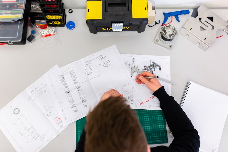
Veterinary orthopedic surgeon Dr. Sebastian uses Bambu Lab H2D to print exact bone replicas for pre-surgical rehearsal, reducing surgery time and improving outcomes for animal patients.
Veterinary orthopedic surgery presents a unique challenge: the patient cannot describe their symptoms, and the anatomical variability between breeds is enormous. A Great Dane femur and a Dachshund femur are not just different in size—they are structurally different in ways that make standardized implants problematic.
Dr. Sebastian, a Doctor of Veterinary Medicine specializing in complex surgical procedures, has developed a solution: using 3D printed bone models for surgical rehearsal before ever touching the actual patient.
The Problem With Traditional 3D Printing
While 3D printing had already become central to Dr. Sebastian's workflow, the technology came with real limitations. Resin printers offered good precision, but hollow bone models required internal support structures that became permanently trapped inside the medullary cavity. The result was anatomically compromised models that were harder to use during surgical rehearsal.
Larger or more complex bones had to be printed in multiple sections and assembled afterward, adding preparation time and introducing inaccuracies. Additionally, the resin printing process required toxic substances during post-processing and a dedicated air treatment system.
Enter Bambu Lab H2D
Bambu Lab sponsored Dr. Sebastian with an H2D—a dual-nozzle, large-format 3D printer. The dual-head system enabled printing with dissolvable support materials, while the generous build volume allowed full-scale bone models to be produced in a single run.
Dissolvable supports changed the game for internal structures. After printing, the supports dissolve away completely, leaving clean medullary cavities that closely match real anatomy—no remnants, no compromises.
The dual-material capability added another layer of utility. Dr. Sebastian can now print combined models showing both the bone and the implant together—exactly as they would appear post-surgery. He can drive bone screws directly into these combined models, simulating the optimal surgical approach before the patient is ever on the table.
Results
The impact was immediate. Anatomically faithful models made surgical rehearsal more straightforward and more useful. For the majority of animal bone cases, models could now be printed as a single piece—eliminating assembly steps and reducing post-processing time.
Dr. Sebastian plans to standardize 3D printing across his practice and build a comprehensive anatomy and lesion model library for team training and intern education.

Comments (0)
No comments yet. Be the first!
Leave a Comment